Taxes included.
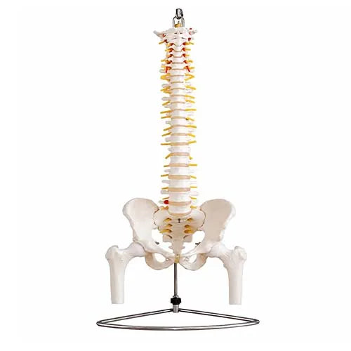
The Spine Model with Pelvis and Femur Heads is a detailed anatomical teaching model designed to help students, educators and healthcare professionals understand the relationship between the vertebral column, pelvis and hip structures. Its flexible spinal construction allows basic demonstrations of spinal movement, posture and alignment.
The model includes a complete pelvis, movable femur heads, occipital plate, visible spinal nerve exits and cervical vertebral artery. It also features an L3-L4 disc prolapse, making it useful for demonstrating common spinal anatomy and explaining disc-related conditions in an educational setting.
With its combination of flexible mounting and detailed anatomical structures, this spine model can be used in medical colleges, physiotherapy institutes, anatomy classrooms, orthopedic and spine clinics, hospitals and patient education environments.
Why Choose This Spine Model?
- Detailed representation of the human vertebral column
- Complete pelvis included
- Movable femur heads for hip-joint relationship demonstration
- Flexible mounting throughout the spine
- L3-L4 disc prolapse represented
- Visible spinal nerve exits
- Detailed cervical vertebral artery
- Occipital plate included
- Mounted on a stable stand
- Useful for classroom and clinical demonstrations
- Suitable for medical and physiotherapy education
- Helpful for patient counselling and anatomy explanation
The combination of these structures is consistent with established flexible spine teaching models used for anatomical demonstration.
Key Features
Flexible Spinal Column
The flexible mounting allows the spine to be positioned and moved for basic demonstrations of spinal alignment and movement.
Complete Pelvis
The included male pelvis helps learners understand the anatomical relationship between the lower spine and pelvic structure.
Movable Femur Heads
Movably mounted femur heads provide a visual reference for explaining the relationship between the pelvis and hip joints.
L3-L4 Disc Prolapse
The model includes an L3-L4 disc prolapse that can be used to explain lumbar disc anatomy and related educational concepts.
Spinal Nerve Exits
Visible nerve exits help demonstrate the pathways where spinal nerves emerge from the vertebral column.
Cervical Vertebral Artery
The detailed cervical vertebral artery provides an additional reference for teaching structures around the upper spine.
Occipital Plate
The occipital plate helps demonstrate the connection between the skull base and upper cervical spine.
Stable Stand
The mounted design makes the model suitable for classroom demonstrations, clinical display and patient education.
Benefits
- Makes complex spinal anatomy easier to visualise
- Supports hands-on anatomy education
- Helps explain the relationship between spine, pelvis and hips
- Useful for demonstrating spinal posture and basic movement
- Supports teaching of lumbar disc anatomy
- Helps explain spinal nerve exit locations
- Provides a practical visual aid for physiotherapy education
- Useful for orthopedic and spine-related patient counselling
- Suitable for repeated classroom demonstrations
- Provides multiple anatomical structures in a single teaching model
Technical Specifications
| Specification |
Details |
| Product Type |
Anatomical Spine Model |
| Model |
Spine with Pelvis and Femur Heads |
| Anatomy Covered |
Vertebral Column, Pelvis, Femur Heads, Occipital Plate, Nerve Exits and Vertebral Artery |
| Pelvis |
Male Pelvis |
| Femur Heads |
Movably Mounted |
| Spine Mounting |
Fully Flexible |
| Disc Feature |
L3-L4 Disc Prolapse |
| Nerve Feature |
Visible Spinal Nerve Exits |
| Artery Feature |
Cervical Vertebral Artery |
| Occipital Plate |
Included |
| Mounting |
Stable Stand |
| Intended Use |
Medical Training and Anatomy Education |
| Suitable For |
Medical Colleges, Physiotherapy Institutes, Clinics, Hospitals and Anatomy Labs |
| Material |
As per manufacturer specifications |
| Model Height |
As per manufacturer specifications |
| Stand Height |
As per manufacturer specifications |
| Weight |
As per manufacturer specifications |
| Warranty |
As per manufacturer specifications |
Frequently Asked Questions
1. What is the Spine Model with Pelvis and Femur Heads used for?
It is designed for anatomy teaching, medical education, physiotherapy training, clinical demonstrations and patient counselling.
2. Does the model include a pelvis?
Yes. The model includes a male pelvis as part of the complete anatomical structure.
3. Are the femur heads movable?
Yes. The femur heads are movably mounted to help demonstrate the relationship between the pelvis and hip joints.
4. Is the spine flexible?
Yes. The MeddeyGo listing specifies flexible mounting throughout the spine, allowing basic movement and alignment demonstrations.
5. Does the spine model show disc prolapse?
Yes. It features an L3-L4 disc prolapse for educational demonstration of lumbar disc anatomy and related conditions.
6. Does it show spinal nerves?
Yes. Visible spinal nerve exits are included to help demonstrate where spinal nerves emerge from the vertebral column.
7. Does the model include the vertebral artery?
Yes. It includes a detailed cervical vertebral artery for anatomy teaching.
8. Is this suitable for physiotherapy institutes?
Yes. The model is suitable for physiotherapy education, anatomy teaching and related clinical demonstrations.
9. Can this model be used for patient counselling?
Yes. Its visible anatomical structures can help healthcare professionals explain spinal, pelvic and hip anatomy in a more visual and understandable manner.
10. Is this a diagnostic medical device?
No. It is an anatomical education and demonstration model and should not be used as a diagnostic device.
Explore More Physiotherapy Machine on medin